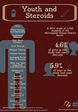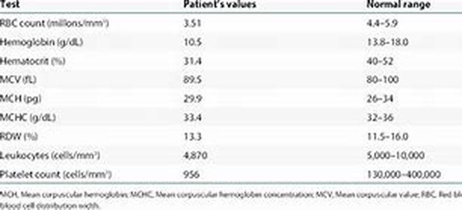
A Hematology Atlas Is A Collection Of Tables, Charts, And Pictures That Deals Primarily With The Morphology Of Blood Cells. Hematology Is That Branch Of Medicine That Deals With The Study Of Blood Cells, Blood Forming Organs, And Blood Diseases. A Specialist In Hematology Is Called A Hematologist And His Work May Range From Laboratory Management And Research To Diagnosis And Treatment Of Disorders Of The Blood. Not Only Hematologists Will Find Some Use From A Hematology Atlas. Laboratory Technicians, Medical School Students, Biologists And Professionals Working In The Field Of Morphology Of Blood Cells May Also Require A Hematology Atlas In Performing Their Responsibilities.The Pictures Contained In A Hematology Atlas Are Magnified With The Use Of Photomicroscopes And Scaled Up For Use As Photographic Sequences. They Show Normal Cells From Blood And Bone Marrow As Well As Blood Cells That Are Affected With A Substantial Variety Of Hematology Diseases. Prior To Capturing The Blood Cells Into Digital Image, The First Thing That The Creator Of The Hematology Atlas Has To Do Is To Conduct A Blood Smear Examination. This Will Accurately Assess The Cellular Morphology Of The Blood By Allowing Quantification Of The Different Types Of Leukocytes And Estimation Of The Platelet Count. In Addition, Blood Smear Examination Images In The Hematology Atlas Will Help Ensure The Detection Of Any Morphological Abnormalities That May Be Indicators Of Pathophysiologic Processes. To Take Full Advantage Of The Value Of Blood Smear Examinations In The Detection Of Hematology Diseases, The Process Requires A Well-prepared And Well-stained Blood Smear As Well As Basic Skills In The Methods Of Assessment. One Of The Most Common Methods Used In Blood Smear Examination For A Hematology Atlas Is The Wedge Smear Preparation. Wedge Smear PreparationThis Is Considered As The Most Convenient, And Hence, Most Commonly Used Technique In The Making Of Blood Smears For Photographic Sequencing In A Hematology Atlas. In Order To Conduct This Technique, The Following Implements Are Required: At Least 3 X 1 Inch (75 X 25 Mm) Clean Glass Slides (use High Quality, Beveled Edge Microscope Slides) Ethylenediaminetetra Acetic Acid (EDTA) Anticoagulated Blood Photomicroscope With Magnifications Of At Least X200First, Allow A Drop (about 3 Mm In Diameter) Of EDTA Anticoagulated Blood To Fall On One End Of The Slide. Then Place The Pusher Slide In Front Of The Drop Of Blood, Making Sure That It Is Positioned At A 30- To 45-degree Angle To The Smear Slide. Pull The Pusher Slide Towards The Drop Of Blood And Hold It In That Position Until The Blood Spreads Across The Width Of The Slide. Finally, Quickly And Smoothly Push Forward To The End Of The Smear Slide. In So Doing, A Wedge Smear Is Created. Capture The Image With The Use Of The Photomicroscope And Magnify For Use In The Hematology Atlas.